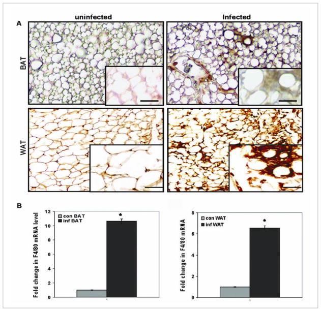
Fig. 2. Macrophages in adipose tissue.
Immunohistochemical analysis using antibody against ionized calcium-binding adaptor molecule 1 (Iba1) of adipose tissue obtained from mice. (A) Brown adipose tissue (BAT), obtained from the interscapluar region and white adipose tissue obtained from the subcutaneous region (WAT). Note the increase in macrophages in the infected adipose tissue. (B) Macrophage-specific F4/80 messenger RNA (mRNA) demonstrating the increase in macrophages in both BAT and WAT, as determined by real-time quantitative polymerase chain reaction. Con=control; Inf=infected (reproduced from Nagajyothi et al. Journal of Infectious Disease [45] with permission from the Journal and the Oxford University Press).

