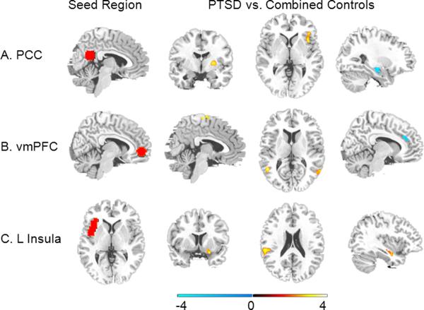
Figure 2. Group Comparison.
(A) Compared to controls, PTSD participants showed greater connectivity between posterior cingulate cortex (PCC; seed region shown in A) and right putamen (column 2; y = −4) and right insula (column 3; z = 2) and a trend for less connectivity between PCC seed and left hippocampus (column 4; x = −27). (B) Compared to controls, PTSD participants showed greater connectivity between ventromedial prefrontal cortex (vmPFC; seed region shown in B) and supplementary motor area (column 2; x = 5) and bilateral superior temporal sulcus (column 3; z = 11) and less connectivity between vmPFC seed and rostral anterior cingulate cortex (column 4; x = −10). (C) Compared to controls, PTSD participants showed greater connectivity between left anterior insula (=seed region shown in C) and right amygdala (column 2; y = 8), left peri-insula (column 3; z = 21) and right hippocampus (column 4; x = 37).

