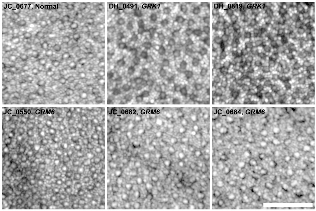
Figure 5.
Images of the perifoveal photoreceptor mosaic obtained with an adaptive optics scanning light ophthalmoscope for 3 patients with complete congenital stationary night blindness caused by GRM6 mutations, 2 brothers with Oguchi disease caused by GRK1 mutations, and 1 normal control. Images are displayed with logarithmic gray scale to facilitate visualization of the rod and cone mosaics. All patients had a complete rod mosaic with a coarser cone mosaic interleaved amongst the rods. Scale bar is 50µm.

