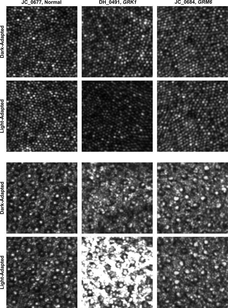
Figure 6.
Imaging the photoreceptor mosaic under different adaptation states in 1 patient with complete congenital stationary night blindness caused by GRM6 mutations, 1 patient with Oguchi disease caused by GRK1 mutations, and 1 normal control. Images are from the parafoveal location (top set) or the perifoveal location (bottom set). The top row in each set shows images immediately after removal from a period of 2 hours of dark adaptation. The bottom row in each set shows images of the exact same patches of retina after about 30–40 minutes into the imaging session, we thus consider these images to be collected under a light adapted state, at least relative to those in the top rows. Each patch of retina is 82 × 82µm, no scale bar is given so as not to obscure any of the cones or rods. The perifoveal rod images of the Oguchi disease were the only ones to show an increase in intensity in the light-versus dark-adapted conditions.

