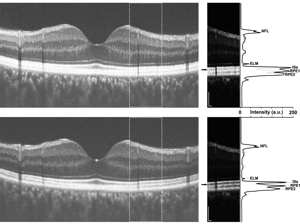
Figure 8.
High-resolution optical coherence tomography line scans (Bioptigen, Inc., Durham, North Carolina, USA) acquired along the vertical meridian of a patient with Oguchi disease caused by GRK1 mutations (DH_0491). Images were acquired under light-adapted (top) or dark-adapted (bottom) conditions. The horizontal line scans on the left are an average of 40 individual B-scans, and are displayed in the conventional logarithmic gray scale. The outer retinal layers are difficult to discriminate in the light-adapted image (top). The inset panels on the right are cropped sections (indicated by the dashed boxes in the larger line scans), displayed in a linear gray scale. Small black arrows indicate the portion of the scan corresponding to the photoreceptor outer segments, clearly visible in both light-adapted (top) and dark-adapted (bottom) conditions. Longitudinal reflectivity profile taken through the center of each inset panel is plotted along the right side of the figure as a solid black line, and demonstrate the presence of the four primary outer retinal layers (external limiting membrane, ELM; inner segment ellipsoid, ISe; and 2 layers corresponding to the retinal pigment epithelium (RPE), RPE1 and RPE2) under both adaptation conditions. The thin gray line on each plot is the longitudinal reflectivity profile from the other imaging condition, allowing comparison of layer position and contrast under the two adaptation conditions. The Michelson contrast of the outer segment layer (the hyporeflective band between the ISe and RPE1) is higher (48%) compared to that in the light-adapted images (12%). Also apparent from this analysis is that the outer segment length (approximated by the distance between ISe and RPE1) was not different between the two adaptation conditions. Scale bar is 100µm.

