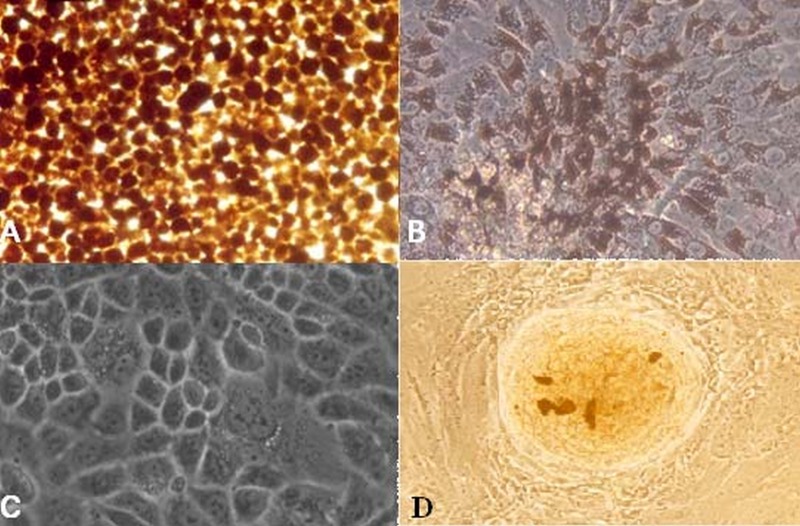
Figure 1.
(A) Retinal pigment epithelial (RPE) cells immediately after separation from a globe. (B) RPE cell culture after one week shows pigment dispersed on the cell surface. (C) Culture after 3 weeks revealed that RPE cells completely covered the floor of the container, retained the appearance of epithelial tissue and lost their pigment to some extent. (D) The appearance of a spheroid colony. The cells were visualized using Zeiss Axiovert 405M inverted microscope (magnification: 10×20 for A, B and D and 10×32 for C).

