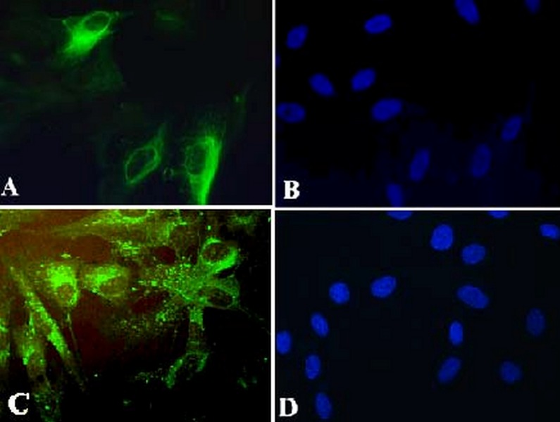
Figure 5.
(A) Retinal pigment epithelial (RPE) cells stained green with cytokeratine antibody.(B) Nuclei of RPE cells are stained blue using 4,6-diamidino-2-phenyindole dihydrocholoride (DAPI). (C) RPE cells are stained green using RPE 65 antibody. (D) Nuclei of the RPE cells stained blue with DAPI (magnification: 8×40).

