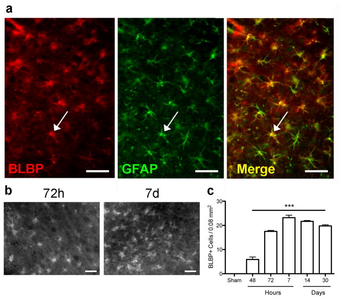
Figure 4.
The number of BLBP immunoreactive cells increases at 48h following MCAO and remains increased through 30 days. (a) Fluorescence micrographs show that BLBP colocalizes with GFAP at 72h following MCAO. Arrow indicates a single cell among the many showing colocalization, yellow on the merged image. (b) Micrographs of BLBP+ cells 72h and 7d after MCAO. (c) Quantification of BLBP-positive cells in the cortical penumbra, showing a significant increase at 48h that remains elevated through 30d. ***p<0.001 versus sham. Scale bar = 50 μm.

