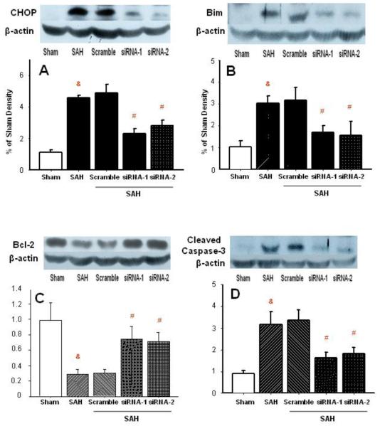
Figure 1.
Western blot analysis of apoptotic proteins in the vascular tissues of basilar artery. CHOP protein was effectively eliminated with siRNA (panel A). Suppressed bim protein level accompanied CHOP decrease (panel B). Bcl-2 protein level showed increase in the group treated with CHOP siRNAs (panel C). Reduction of cleaved caspase-3 in BA of rats treated with CHOP siRNAs (panel D). Panels A-D: n=6 in each group (&p<0.05 vs. sham; #p<0.05 vs. SAH, ANOVA).

