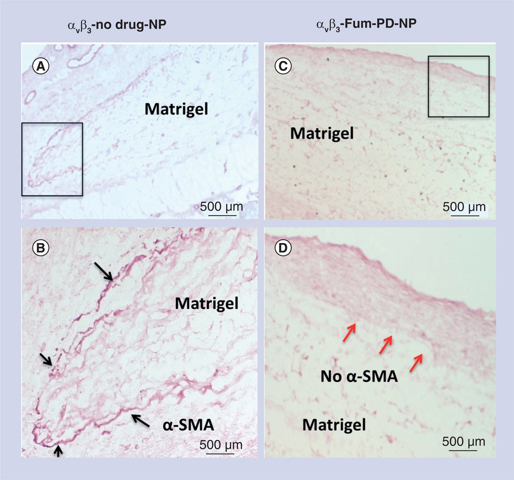
Figure 5. Microscopic examination of FGF Matrigel™ subcutaneous explant in mice following serial injection (intravenous) of αvβ3-targeted no-drug perfluorocarbon nanoparticles (αvβ3-no drug-NP) or αvβ3-targeted fumagillin prodrug perfluorocarbon nanoparticles (αvβ3-Fum-PD-NP).
(A) Low power image of Matrigel™ explant from control animal that was immunostained to identify maturing vessels with α-SMA. Positive staining was appreciated peripherally around the majority of the plug (black box). (B) Enlarged view of boxed region in (A), showing the peripheral α-SMA biomarker. (C) Low power image of Matrigel explant from fumagillin PD-treated animal that was immunostained to identify maturing vessels with α-SMA. Virtually no positive staining was appreciated along the implant periphery, suggesting that the repeated pruning of angiogenic vessels with fumagillin PD impaired the development and progression of mature vessels in contrast with the control. (D) Enlarged view of boxed region in (C), illustrating the lack of peripheral α-SMA biomarker (red arrows).
α-SMA: α-smooth muscle actin; NP: Nanoparticle; PD: Prodrug.

