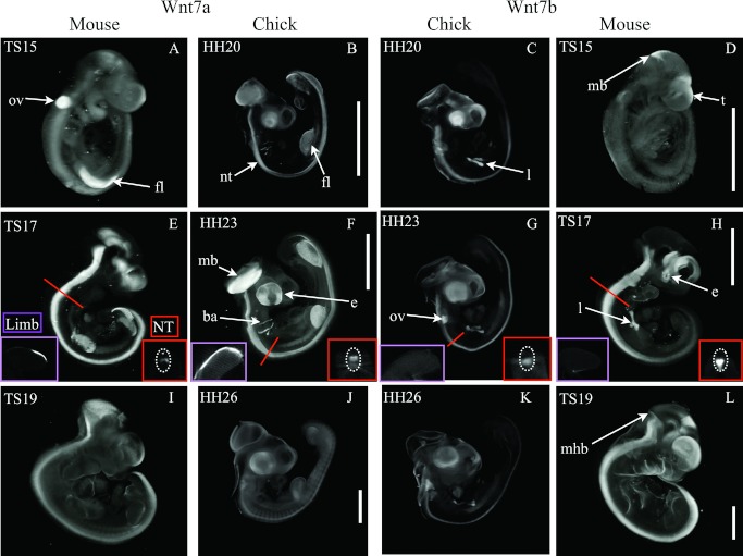
Fig 7.
Wnt7 subfamily gene expression in mouse and chick embryos at stages indicated. Main images are external views (volume rendered) of whole embryos following three-dimensional imaging (OPT): mouse Wnt7a (A, E, and I); chick Wnt7a (B, F, and J); chick Wnt7b (C, G, and K); mouse Wnt7b (D, H, and L). Virtual longitudinal sections through the limb (dorsal up, ventral, down) are inset in (E–H) for the respective genes and stages. Virtual transverse sections through the neural tube at the position indicated are also inset in (E–H). Abbreviations: ba, branchial arch; e, eye; fl, forelimb; l, lung; mb, midbrain; mhb, midbrain-hindbrain boundary; nt, neural tube; ov, otic vesicle. Scale bars indicate 2 mm and are indicative for each stage.

