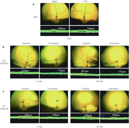
Figure 3.
In vivo assessment of retinal morphology in dogs A3 and A7 at 4 and 18 mpi. (a) Fundus photographs and retinal cross-sectional images obtained from control nonaffected, untreated dog NA1 at 18 months of age. (b) Fundus photographs and retinal cross-sectional images obtained from dog A3 treated with AAV2/5RK.cpde6β at 4 and 18 mpi. (c) Fundus photographs and retinal cross-sectional images obtained from A7 treated with AAV2/8RK.cpde6β at 4 and 18 mpi. Dark circles on fundus photographs schematically represent areas of the treated retinas exposed to recombinant adeno-associated virus (rAAV) vectors. Optical coherence tomography (OCT) scans were acquired on a horizontal line shown on the fundus images (dark-line). The localization and the size of the dark-lines represent the localization and the size of the OCT scans. Retinal thicknesses at the same location were measured using calibrated calipers and indicated on the OCT scan. mpi, months postinjection; µm, micrometers.

