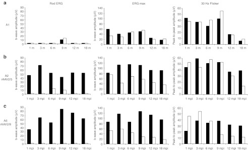
Figure 6.
Kinetics of retinal function recovery in treated dogs A2 and A6. (a) Amplitudes of electroretinography (ERG) responses for control affected, untreated dog A1 from 1 to 18 months of age. (b) Amplitudes of ERG responses for dog A2 treated with AAV2/5RK.cpde6β from 1 to 18 mpi. (c) Amplitudes of ERG responses for dog A6 treated with AAV2/8RK.cpde6β from 1 to 18 mpi. The left and middle panels show scotopic rod and mixed cone-rod-mediated b-wave amplitudes, respectively. The right panel shows photopic 30 Hz flicker amplitude. Right eyes are shown in dark, left eyes in white. AAV, adeno-associated virus; mpi, month(s) postinjection.

