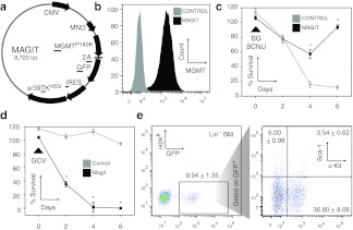
Figure 1.
In vitro validation of transduction, expression, and function of MGMTP140K-2A-GFP-IRES-TK (MAGIT) lentivirus. (a) Map of MAGIT tricistronic plasmid co-expressing MGMTP140K, enhanced green fluorescent protein (eGFP) linked by a 2A sequence, and followed by TKHSV separated by an internal ribosome entry site (IRES). (b) 293T cell line was transduced with MAGIT lentivirus and intracellular MGMTP140K and GFP expression analyzed by flow cytometry. The proportion of cells shown expressing intracellular MGMTP140K and GFP expression was >90% positive cells as compared with untransduced cells. In all histograms the y-axis represents cell counts. (c) The same cell population in b was exposed to BG followed by BCNU, separated by 1 hour. Survival as a percentage of untreated cells was determined in triplicates at 2-day intervals. (d) Simultaneous to c, MAGIT-transduced cells were exposed to Ganciclovir (GCV) and monitored similarly. (e) Lineage-depleted bone marrow from C57Bl/6 mice was transduced with MAGIT lentivirus and the levels of GFP, Sca-1, and c-Kit expression determined by flow cytometry 48 hours following transduction. The dot plot showing Sca-1 and c-Kit expression is gated to include only GFP+ cells. These data are representative of at least three independent experiments and any differences with P < 0.05 are indicated with an asterisk (*). Gray: control-untransduced cells; black: MAGIT-transduced cells. BCNU, 1,3-bis(2-chloroethyl)nitrosourea; BG, benzylguanine; CMV, cytomegalovirus.

