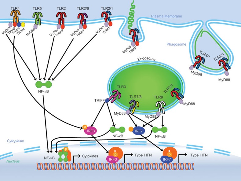
Figure 1.
TLR signaling overview: the TLR family can be subdivided into two categories. TLR1, TLR2, TLR4, TLR5 and TLR6 are located primarily on the cell surface. Conversely, TLR3, TLR7, TLR8 and TLR9 are located within intracellular vesicles. Furthermore, TLRs can be found as homo or heterodimers (such as TLR1/2 and TLR2/6). Upon recognition of a PAMP, the TLR changes its conformation and recruits adapter proteins such as TRIF, TRAM, MyD88 and TIRAP. The cell surface TLRs, including the heterodimers TLR1/2 and TLR2/6, recruit MyD88 and TIRAP adaptor proteins to signal the NF-κb pathway to produce NF-κb-mediated cytokines. Additionally, TLR4 uses the TRIF and TRAM proteins to produce type I IFNs through the IRF3 pathway. Of the endosomal TLRs, TLR8 and TLR9 utilize adaptor protein MyD88 to signal through NF-κb and IRF7 pathways. Finally, endosomal TLR3 enlists the TRIF adaptor to create both type I IFNs as well as NF-κb cytokines through IRF3 and NF-κb pathways. The model depicts internalization of an extracellular pathogen, Borrelia burgdorferi (Bb), which is internalized into the phagosome where TLR2 and TLR8 signalling occurs. Phagocytosis of live Bb, induces transcription of IFN-β through IRF-7, a phenomenon entirely dependent on the availability of TLR8, while induction of NF-κb-dependent cytokines is due to a cooperative action of TLR2 and TLR8.10 IFN, interferon; IRF, IFN regulatory factor; PAMP, pathogen-associated molecular pattern; NF, nuclear factor; TLR, Toll-like receptor.

