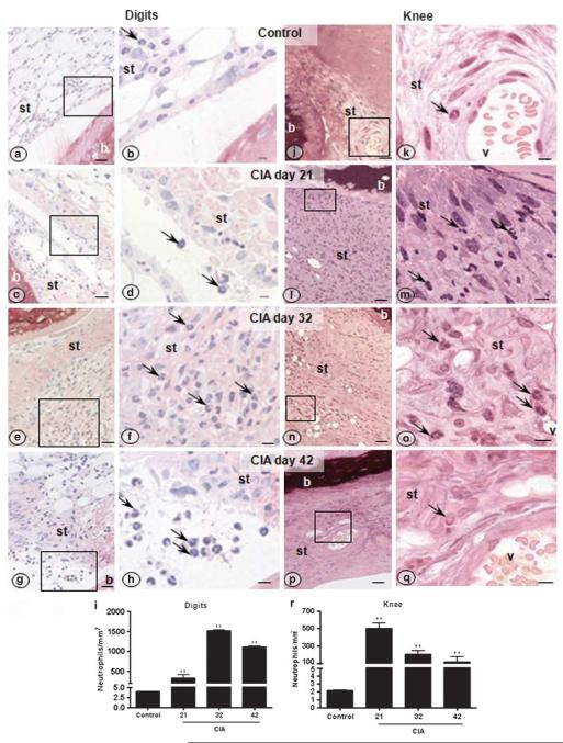
Figure 2.
Neutrophils migration after CIA within the synovial tissue of digits and knees joints. Synovial tissue of the digits (a–i) and knees (j–r) displayed an evident accumulation of transmigrated neutrophils (arrows) on days 21, 32 and 42 after collagen immunization. Data were analyzed using one way ANOVA followed by Dunnett’s post hoc test. Results are expressed as the mean±s.e.m. taking a P-value less than 0.05 as significant (n = 5 animals per group). **P<0.01 vs control group; P<0.001 vs digits. Picrosirius and H&E-stain; scale bars: (a, c, e, g, j, l, n, p) 20 μm; (b, d, f, h, k, m, o, q) 5 μm. v, vessel; st, synovial tissue; b, bone.

