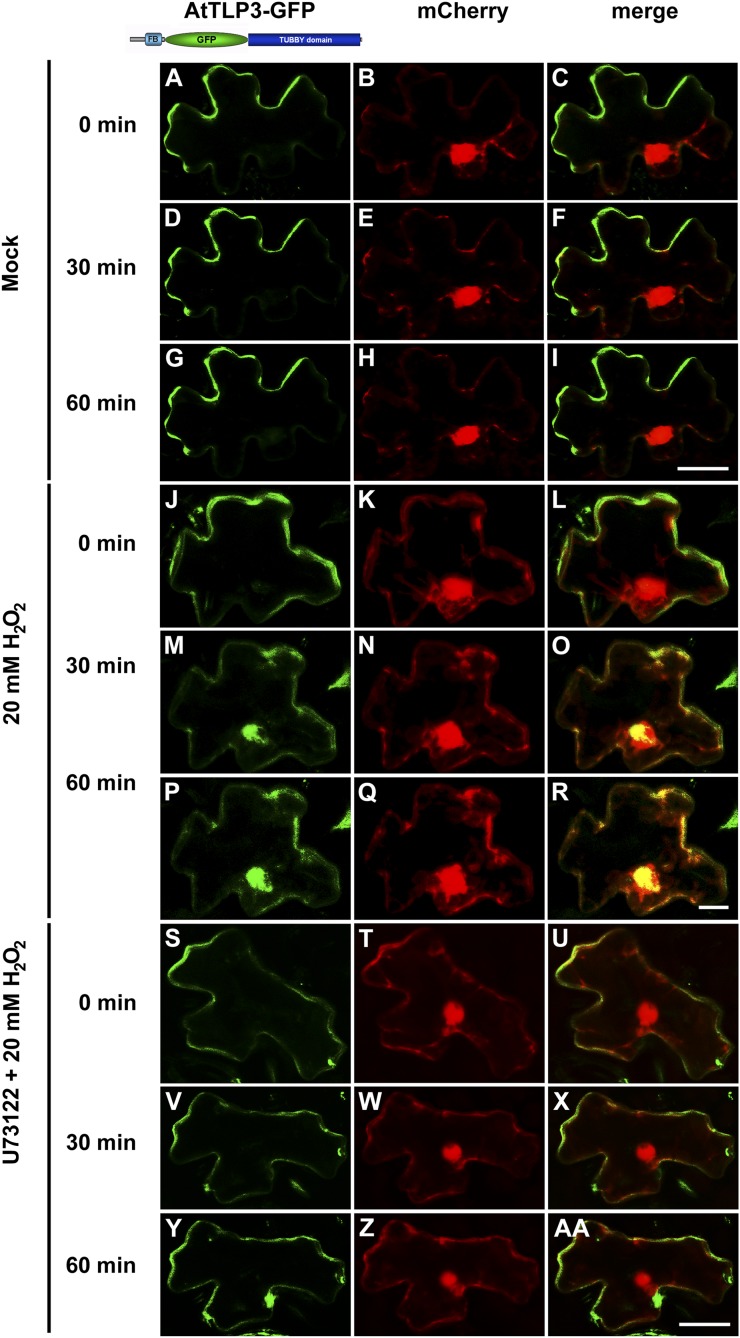
Figure 9.
Phospholipase C releases full-length AtTLP3-GFP from the PM when heterologously expressed in N. benthamiana. A, D, G, J, M, P, S, V, Y, N. benthamiana leaf cells were transformed with full-length AtTLP3-GFP. B, E, H, K, N, Q, T, W, Z, N. benthamiana leaf cells were transformed the cytosolic and nucleoplasmic marker mCherry. C, F, I, L, O, R, U, X, AA, Merged images indicate the subcellular localization of full-length AtTLP3-GFP. Yellow color indicates the colocalization of green- and red-fluorescing proteins. A to I, Full-length AtTLP3 fused to GFP displayed PM localization over time. J to R, Application of 20 mm H2O2 results in the relocalization of full-length AtTLP3-GFP from the PM to the cytosol and nucleus. J to L, PM localization of full-length AtTLP3-GFP before treatment (0 min). M to O, Cytosolic and nucleoplasmic localization of full-length AtTLP3-GFP at 30 min after application of 20 mm H2O2. P to R, Cytosolic and nucleoplasmic localization of full-length AtTLP3-GFP at 60 min after application of 20 mm H2O2. S to AA, Pretreatment with the phospholipase C inhibitor U73122 inhibits H2O2-triggered relocalization of full-length AtTLP3-GFP from the PM to the cytosol and nucleus. Treated cells were observed at 0 min (S–U), 30 min (V–X), and 60 min (Y–AA) after H2O2 application. Experiments were repeated three times with similar results. Bars = 20 µm.

