
Figure 1.
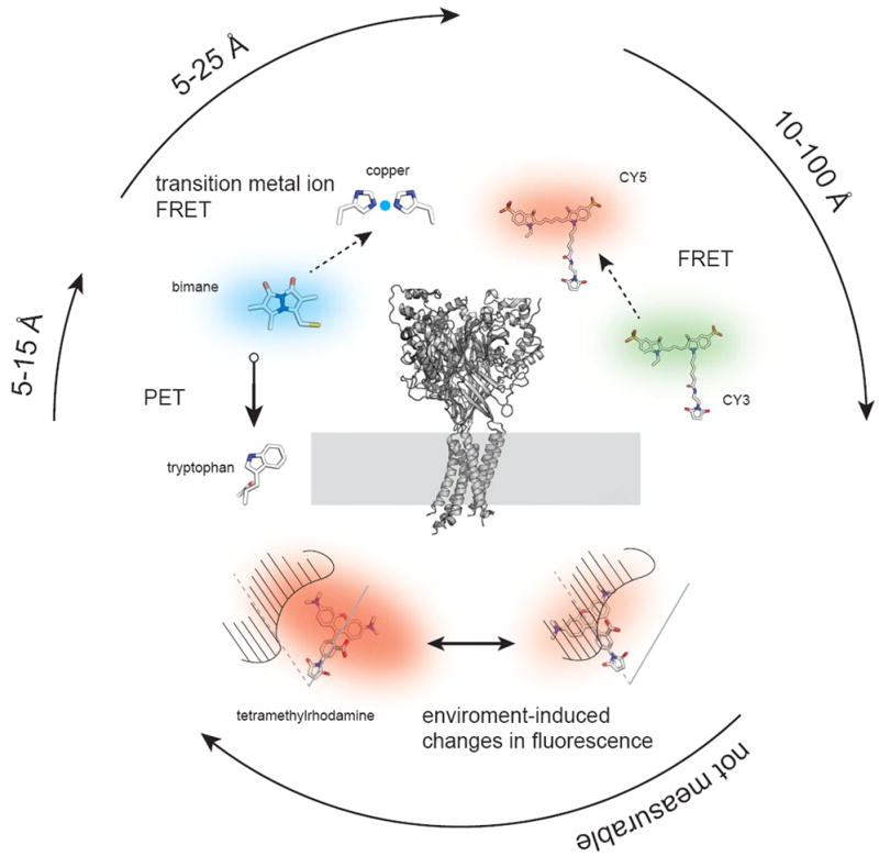
Fluorescent methods used to explore a membrane protein structure. In the center is the crystal structure of the acid sensing ion channel ASICS1 (PDB 2QTS) positioned with its transmembrane domains in a membrane [61]. Surrounding the structure are example fluorescent methods including FRET between the two fluorescent dyes Cy3 and Cy5, transition metal ion FRET between the dye bimane and a di-histidine bound metal, photo-induced electron transfer (PET) between bimane and a tryptophan, and a tetramethyl-rhodamine fluorophore undergoing an environment-induced fluorescent change. The approximate distance scales that each technique works over are indicated by the surrounding arrows.