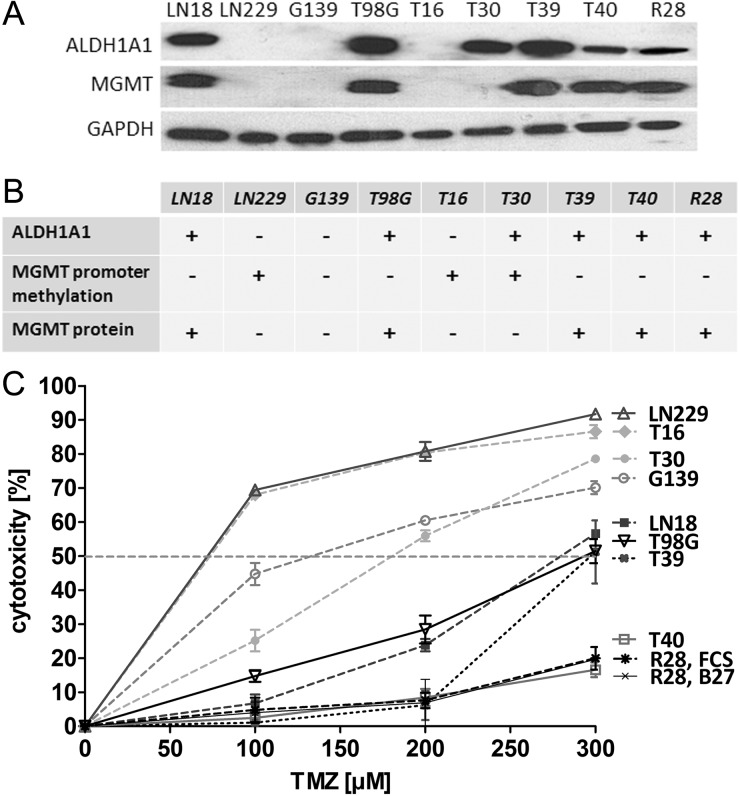
Fig. 1.
ALDH1A1 expression, MGMT status, and response to treatment with TMZ. (A) Western blot analysis showed a high level of ALDH1A1 in LN18, T30, T39, T40, T98G, and R28 cells. The cell lines LN229, G139, and T16 were negative for ALDH1A1. MGMT protein expression was detected in LN18, T98G, T39, T40, and R28 cells. (B) No relevant MGMT promoter methylation was found in LN18, T98G, G139, T39, T40, or R28 (MGMT+). The MGMT promoter of T16, T30, and LN229 was methylated to 60%, 30%, and 100%, respectively (MGMT–).The MGMT promoter of G139 cells was completely unmethylated, but no expression of MGMT protein was detected (MGMT–*). (C) Response of GBM cell lines to treatment with TMZ for 7 days was assessed by metabolic MTT assay.

