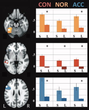
Figure 4.

Region of interest analyses. Left: Regions (white spheres). Left fusiform (top, yellow); middle temporal (middle, red); and inferior frontal (bottom, blue) gyri. Colored areas indicate greater within‐structure activation for fluent sentence reading > rest. Right: Activation for [fluent (S)entence reading > rest] vs [(L)etter reading > rest]. CON = constrained; NOR = normal; ACC = accelerated. *Significantly different (P<0.05). Axial slices at −20 (fusiform), −2 (middle temporal), and 26 (inferior frontal). [Color figure can be viewed in the online issue, which is available at wileyonlinelibrary.com.]
