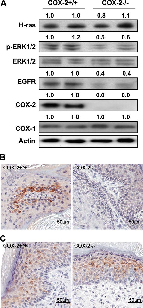
Fig. 4.

Western blotting and immunohistochemistry of COX-2+/+ and COX-2−/− papillomas. (A) Protein lysates were prepared from two COX-2+/+ papillomas and two COX-2−/− papillomas and analyzed for H-ras, p-ERK1/2, total ERK1/2, EGFR, COX-2 and COX-1 by western blotting (30 µg per lane loaded). The numbers above each lane represent densitometry readings. (B) COX-2 immunostaining of COX-2+/+ papillomas indicated that COX-2 is primarily located in the basal cells and, as expected, COX-2 was not detected in the COX-2−/− papillomas. (C) COX-1 immunostaining indicated that the COX-2+/+ and COX-2−/− papillomas expressed COX-1 in the suprabasal cell layers at comparable levels in agreement with the western blotting results.
