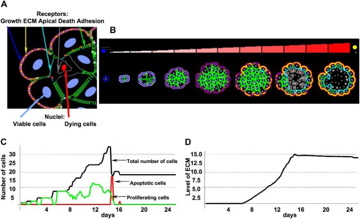
Fig. 2.
(A) Colour-coded cell membrane receptors: cell–cell adhesion (green), cell–ECM adhesion (yellow), apical (cyan), growth (blue), death (grey). (B) A sequence of consecutive stages in the development of a hollow acinus starting from a single cell and ending with a monolayer structure enclosing the hollow lumen. Accumulating ECM proteins are indicated by different shades of red (see colour bar) and the change of receptors from growth (blue) to ECM (yellow). (C) Evolution of different cell subpopulations: proliferating (green), apoptotic (red) and total (black) over the time course of acinar formation. (D) Dynamics of ECM secretion and accumulation along the perimeter of an emerging acinus. Both data sets correspond to the morphologies shown in panel (B). Model parameters: growth threshold: 15%, death threshold: 25%, ECM threshold: 15×103κ1.

