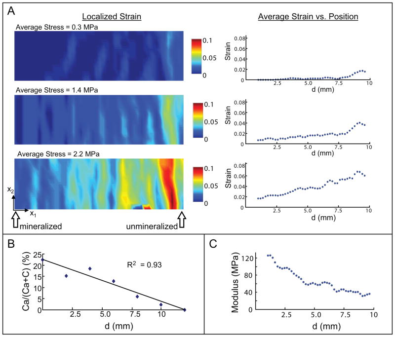
Figure 3.
Nanofiber scaffolds were synthesized with gradations in mineral to mimic the natural tendon-to-bone insertion. There was a gradation in mechanical properties along the length of the scaffolds (a representative PLGA scaffold is shown). (A) The strains in the x1 direction for three values of stress are shown. Localized strains are shown on the left and average strains are shown on the right. Strain increased with increasing stress and was highest on the unmineralized side of the scaffold. (B) There was a linear decrease in calcium phosphate along the length of the scaffold. (C) Young’s modulus decreased with decreasing calcium phosphate content. [Reproduced, with permission, from: Li X, Xie J, Lipner J, Yuan X, Thomopoulos S, Xia Y. Nanofiber scaffolds with gradations in mineral content for mimicking the tendon-to-bone insertion site. Nano Letters. 2009;9(7):2763–8.]

