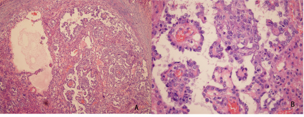
Figure 4.
Histopathology of the resected specimen.A. The resected specimen was a malignant tumor composed of neoplastic cells proliferating in a microcystic or reticular growth pattern. B. Neoplastic cells with highly atypical middle-or large-sized nuclei and Schiller-Duval bodies partially mimicking papillary structures

