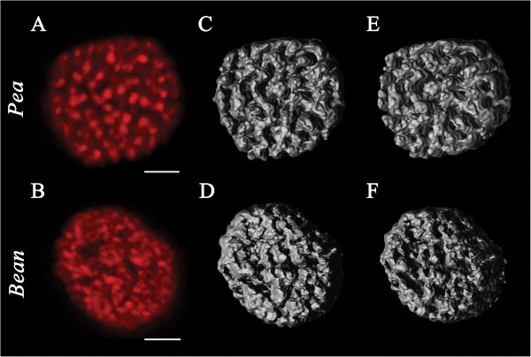
Figure 1.
Chlorophyll fluorescence of intact chloroplasts revealed by CLSM and 3D reconstruction of chloroplast structure. The intact chloroplasts of pea (A) and bean (B) were incubated in isoosmotic medium containing 15 mM NaCl and 4 mM MgCl2. Each red image presents the maximum intensity projection of deconvolved stack of CLSM images. Bar = 2 μm. Images are representative for at least 20 independent experiments. Grey images represent 3D models of intact pea (C, E) and bean chloroplasts (D, F) created after deconvolution. Face (C, D) and side view (E, F) of 3D chloroplast models is shown. Each image is a representative of at least 10 independent experiments.

