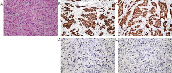
Figure 1.
Immunohistochemical analysis of VEGF-C and VEGF-D expression in primary breast carcinoma. (A) Histopathology by hematoxylin-eosin staining of breast cancer; (B) Strong expression of VEGF-C and (D) negative control in breast cancer; (C) Strong expression of VEGF-D and (E) negative control in breast cancer. Diffuse, strong positive VEGF-C (B) and VEGF-D (C) immunostaining was mainly observed in the cytoplasm of breast cancer cells (magnification × 400). VEGF-C/D, vascular endothelial growth factor C /D.

