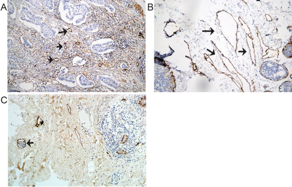
Figure 2.

Immunohistochemical analysis of lymphatic vessels in primary breast carcinoma. (A) Intense, specific D2-40 immunoreactivity was only observed in lymphatic endothelial cells. The intratumoral lymphatic vessels are small, irregular and collapsed (arrow). (B) The peritumoral lymphatic vessels located at the invasive edge of tumors are frequent, often large and dilated (arrow); magnification × 200.(C) Immunohistochemical visualization of invading breast cancer cells in the lymphatic vessels of the peritumoral region of a primary breast carcinoma. The black arrow indicates lymphatic vessel invasion (LVI); magnification × 200.
