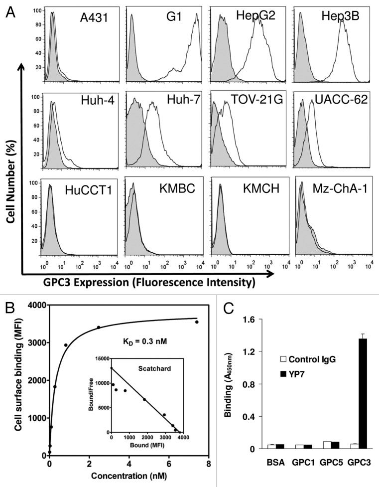
Figure 2. Characterization of YP7 binding properties. (A) Flow cytometry analysis using a panel of endogenous GPC3–expressed human cell lines. Four HCC (HepG2, Hep3B, Huh4, Huh7), four CCA (HuCCT1, KMBC, KMCH and Mz-ChA-1), one ovarian CCC (TOV-21G) and one melanoma (UACC-62) cell lines were shown. Filled peak: isotype control. (B) Binding affinity measurement of YP7 against cell surface-associated GPC3. MFI: mean fluorescence intensity. The Scatchard plot and the KD value were calculated using Prism. (C) Binding of YP7 on recombinant GPC1, GPC3 and GPC5 proteins.

An official website of the United States government
Here's how you know
Official websites use .gov
A
.gov website belongs to an official
government organization in the United States.
Secure .gov websites use HTTPS
A lock (
) or https:// means you've safely
connected to the .gov website. Share sensitive
information only on official, secure websites.
