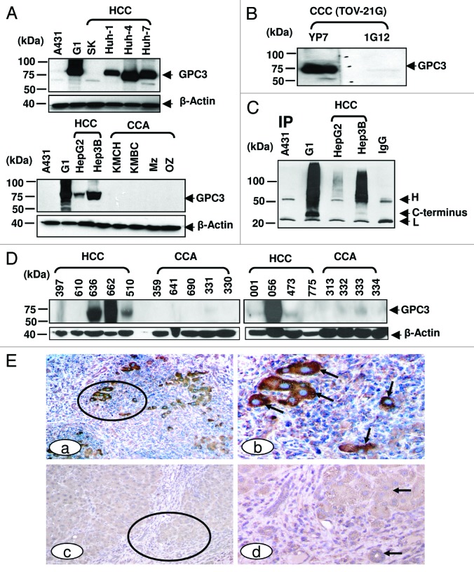
Figure 3. GPC3 protein expression in liver cancer. (A) GPC3 expression in liver cancer cell lines detected in western blotting. HCC: hepatocellular carcinoma; CCA: cholangiocarcinoma. 1G12: a commercial GPC3 mAb. Mz: Mz-ChA-1; SK: SK-Hep-1. (B) western blots of ovarian clear cell carcinoma (CCC). TOV-21G: ovarian CCC. (C) Soluble GPC3 protein immunoprecipitated from culture supernatant and analyzed by western blotting. YP7 was used to pull down soluble GPC3 protein in culture supernatant. H: mouse IgG heavy chain; L: mouse IgG light chain. C-terminus: C-terminal subunit (~30 kDa). IP: immunoprecipitation. (D) GPC3 expression analyzed by western blotting in nine HCC and nine CCA primary liver tumor tissues. (E) Immunohistochemistry of GPC3 in HCC tissues. Circles identify tumor nests (a, c) with or without distinct GPC3 positivity at a low magnification. Arrows identify tumor (b, d) with or without GPC3 positivity at a higher magnification. a, c: 100X. b and d: a higher magnification (400X) of a and c, respectively.

An official website of the United States government
Here's how you know
Official websites use .gov
A
.gov website belongs to an official
government organization in the United States.
Secure .gov websites use HTTPS
A lock (
) or https:// means you've safely
connected to the .gov website. Share sensitive
information only on official, secure websites.
