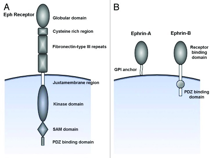
Figure 1. Schematic representation of the structure of Eph receptors and ephrins. (A) Eph receptors are divided into two subclasses based on their sequence homology of the extracellular ephrin-binding globular domain. Eph receptors also have an extracellular cystein rich region followed by two fibronectin-type III repeats. The juxtamembrane domain contains tyrosines that can undergo autophosphrylation, and is followed by a tyrosine kinase domain. The SAM and PDZ binding domains serves as docking sites for interacting proteins and mediators of downstream signaling. (B) Ephrins are subdivided based on how they are linked to the membrane; ephrin-As are linked through a glycosylphosphatidyl anchor, whereas ephrin-Bs contain a transmembrane domain as well as a cytoplasmic tail involved in signaling processes.

An official website of the United States government
Here's how you know
Official websites use .gov
A
.gov website belongs to an official
government organization in the United States.
Secure .gov websites use HTTPS
A lock (
) or https:// means you've safely
connected to the .gov website. Share sensitive
information only on official, secure websites.
