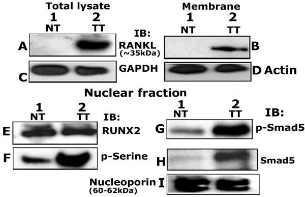
Figure 9.
Western analyses in prostatic normal and tumor lysates. Total cellular (A and C), membrane (B and D) and nuclear (E to I) lysates from normal (NT) and prostatic tumor (TT) tissue (~20 μg protein) were immunoblotted (IB) with a RANKL (A and B), RUNX2 (E), phosphoserine (p-Serine; F), phospho-Smad 5 (p-Smad 5; G) and Smad 5 (H) antibody. Equal loading of the protein was shown in total cellular, membrane and nuclear lysates by relevant immunoblotting analysis with antibodies to GAPDH (C), actin (D) and nucleoporin (I). The results shown are representative of three independent experiments with three different lysates purchased from the vendor.

