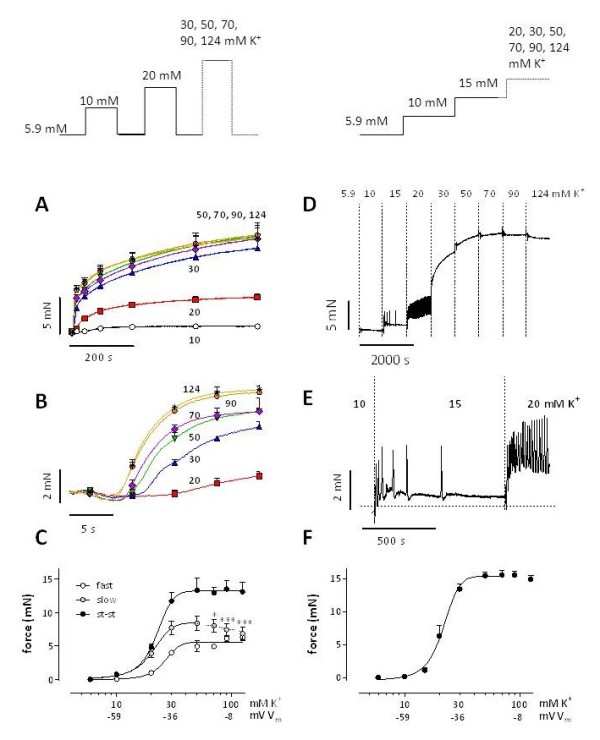
Figure 1.

Isometric contractions by elevation of external K+in mouse aorta. K+ was elevated from 5.9 mM to 10, 20, 30, 50, 70, 90 or 124 mM K+ according to the protocols shown in the top panels. For the repetitive protocol (A-C), traces, shown on condensed (A) and expanded (B) time scales, were analyzed with a bi-exponential function revealing [K+]-force curves for the fast, slow and steady-state (st-st) force components (C). For the cumulative protocol (D-F), D shows a representative example of isometric force elicited by gradual elevations of extracellular K+. E displays the 5.9 to 10, 10 to 15 and 15 to 20 mM K+ depolarizations on an expanded time scale. In F “steady-state” force at each step was plotted in function of [K+]. Results show mean ± s.e.m, n = 4 (A-C) or n = 5 (F). *, ***: P<0.05, 0.001 decrease of slow component amplitude versus maximum at 50 mM K+. The estimated values of Vm at 10, 30 and 100 mM [K+] are indicated. These values are respectively −59, -36 and −8 mV (see Additional file 1).
