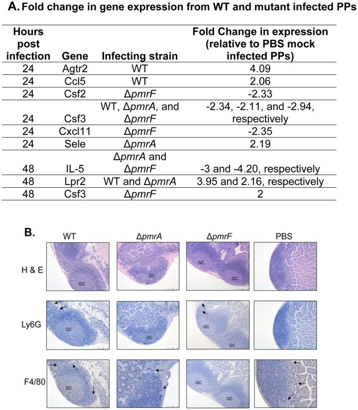
Figure 6. Immune response to oral infection with 108 CFU of WT, ΔpmrA, or ΔpmrF S. Typhimurium.
(A.) Significant changes (2-fold or greater relative to uninfected controls) in immune-related gene expressionfrom mouse Peyer’s patches infected with WT, ΔpmrA, or ΔpmrF S. Typhimurium relative to PBS mock-infected Peyer’s patches. (B.) Hematoxylin and Eosin staining, and immunohistochemical stainging of Peyer’s patches infected with WT, pmrA, pmrF S. Typhimurium, or mock-infected with PBS for 48 hrs. Neutrophils were stained using Ly6G antibodies. Macrophages were stained using F4/80 antibodies. GC- germinal center.

