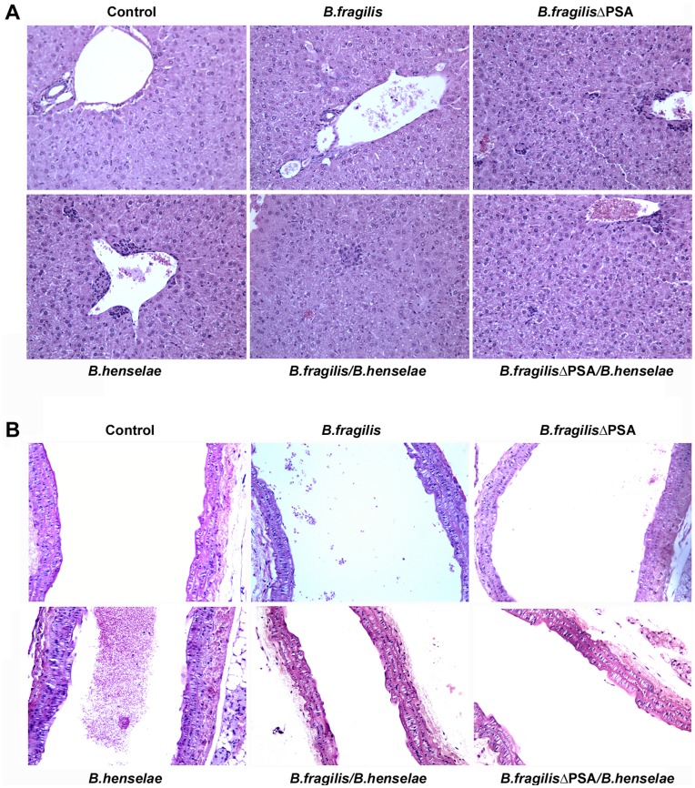
Figure 4. Morphological analysis of murine coinfected tissue by hematoxylin-eosin staining. A.
Representative microscope images of hematoxylin-eosin staining of liver tissues from each group of mice uninfected, infected with B. henselae, B. fragilis and B. fragilis ΔPSA or coinfected, as detailed. Granulomatous inflammatory infiltrates are predominantly evident in the group of mice infected with B. henselae compared to the group infected with B. henselae and B. fragilis ΔPSA and particularly in the mice coinfected with B. henselae and B. fragilis. Uninfected controls are negative, as well as B. fragilis and B. fragilis ΔPSA groups. B. Representative sections stained with hematoxylin-eosin of aortas from all mice groups as described. In the group of mice infected with B. henselae, minimal lesions are observed in the aorta compared to liver tissues.

