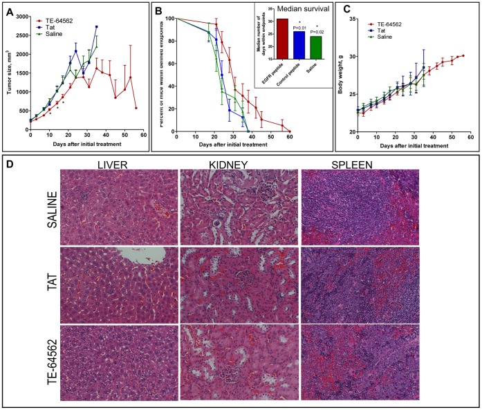
Figure 4. TE-64562 inhibits tumor growth in MDA-MB-231 xenograft tumors and increases survival with no observed toxicity.
(A–C) MDA-MB-231 xenograft tumors were grown in the subcutaneous flank region of nude mice which were treated bi-weekly with the TE-64562 peptide (40 mg/kg; 7 µmol/kg), Tat-peptide (20 mg/kg; 7 µmol/kg) or vehicle (saline), intraperitoneally. (A) The mean tumor size (± standard error of the mean) is plotted over time. The asterisks (*P≤0.0325) indicate that the mean size of the TE-64562 treated tumors is statistically different from the saline- and Tat-treated tumor sizes at that time point. (B) The number of mice within endpoints, as defined by tumor size cutoff, tumor ulceration and body conditioning scoring, at each time point are plotted as a Kaplan and Meier survival curve. (B, inset) The median survival, the number of days at which the fraction of mice within endpoints is equal to 50%, is plotted for each treatment group. The survival curves for the Tat and Saline groups were compared to the survival curve for the TE-64562 group and the P value was derived using the log-rank (Mantel-Cox) test. The asterisks (*) designate a significant difference with the indicated P values. (C) The mean body weight (± standard error of the mean) for each treatment group is plotted over time. (D) After 35 days of dosing, organs were collected and fixed. Representative H&E stained sections from liver, kidney and spleen are shown for each treatment group. Results are representative of two independent studies. Also see Figure S4.

