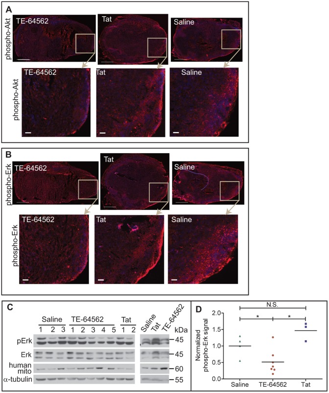
Figure 8. TE-64562 treatment reduces Akt and Erk phosphorylation in MDA-MB-231 xenograft tumor tissue.
(A–B) Nude mice bearing subcutaneous, MDA-MB-231 xenographic tumors were injected with the TE-64562 peptide (40 mg/kg; 7 µmol/kg), Tat-peptide (20 mg/kg; 7 µmol/kg) or vehicle (saline), intraperitoneally for four days, once per day. On the last day, the mice were injected 30 minutes prior to extracting the tumor. Frozen tumor sections were stained for (A) phospho-Akt (S473) or (B) phospho-Erk and counterstained with DAPI. Representative stained tumor sections are shown with the area in the box enlarged in the images below each section. Large scale bars = 500 µm and small scale bars = 50 µm. (C) A ∼1–2 mm cross-sectional slice of the tumor was lysed in RIPA buffer by sonication and the resulting lysates were analyzed by Western blot. Each lane represents a tumor from a different mouse. (D) Western blot data is quantified and plotted. Each treatment group was compared statistically (*P≤0.0364). Error bars represent the standard error of the mean. Also see Figure S6.

