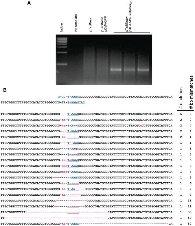
Figure 1. Transposase-mediated excision of the TcBuster transposon in HEK-293 cells.
(a) An agarose gel of the excision PCR. Plasmid DNA was extracted from transfected HEK-293 cells and used as a template for nested PCR to detect the excision of the transposon DNA. Lane 1, 1 kb ladder; lane 2, PCR reaction without any DNA template added; lanes 3–7, PCR on extracts from cells transfected with either 1 µg of the transposon plasmid pTcBNeo (lane 3), 867 ng transposon pTcBNeo and 133 ng pCMVGFP negative control (lane 4), or 867 ng transposon pTcBNeo and 133 ng pXL-CMV-TcBusterCO transposase plasmid (three separate transfections, lanes 5–7). (b) The three PCR bands shown in (a) were gel-purified and TOPO-cloned. Clones were sequenced to determine the exact excision junction. The sequence flanking the transposon in pTcBNeo is shown at the top. The TAAAG homology region is shown in blue. Mismatches are shown in lowercase pink. Dashes are used to maintain alignment and if pink, indicate a missing bp. The sequences are ranked according to (1) incidence (# of clones) followed by (2) number of bp not matching the highest incidence clone (# bp mismatches).

