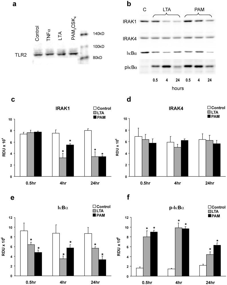
Figure 1. LTA- or PAM-induced activation of the TLR2 pathway in pulmonary microvessel endothelial monolayers (PMEM).
(a) Representative Western blot of TLR2 in PMEM after treatment with vehicle, TNFα 100 ng/mL, LTA from S. aureus and PAM (both 30 µg/mL) for 1 hour, confirming presence of receptors. (b) Representative Western blots of IRAK1, IRAK4, IκBα, and p-IκBαSer32/36 from Control (C) or 0.5, 4 and 24 hour LTA or PAM-treated (both 10 µg/mL) PMEM and (c–f) the blot band densities in Relative Density Units (RDU) for these proteins from all blots of similar treatments. Values represent means ± SEM (N ≥4). * p<0.05 vs. Control.

