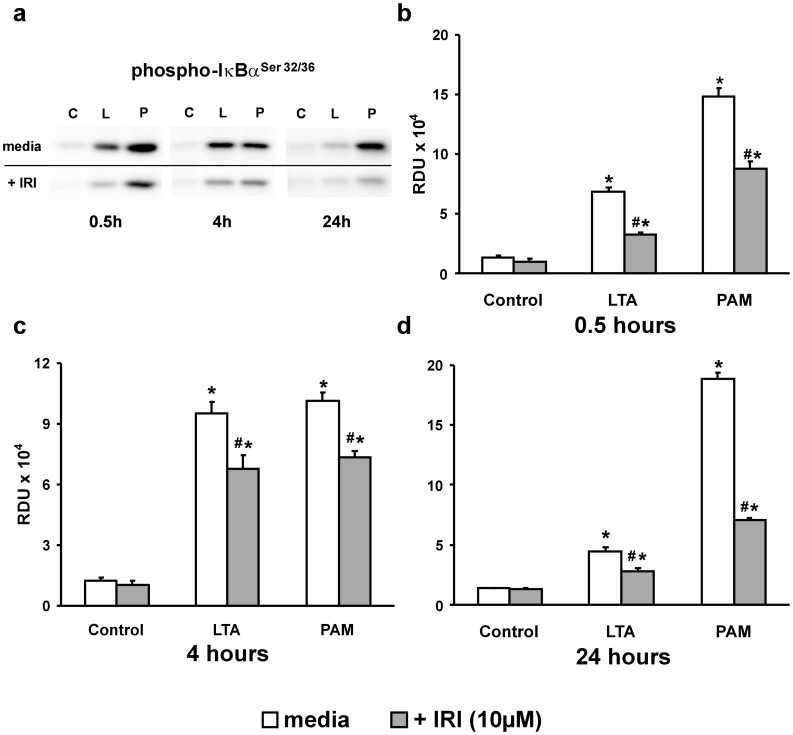
Figure 2. IRAK1/4 inhibition of the TLR2 pathway.
(a) Western blots of p-IκBαSer32/36 from 0.5, 4 and 24 hour Control (C), LTA (L) or PAM (P) -treated (both 10 µg/mL) PMEM in the absence (upper bands) or presence (lower bands) of IRAK1/4 inhibitor (IRI: 10 µM). (c–d) Western blot band densities in Relative Density Units (RDU) for p-IκBαSer32/36 from all blots represented by (a). Values represent means ± SEM (N ≥4). * p<0.003 vs. Control, # p<0.02 vs. TLR2 agonist alone.

