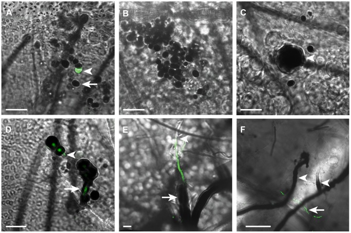
Figure 1. Mosquito melanization response to B. bassiana developmental stages.
Merged fluorescent and bright field images of abdomens dissected from mosquitoes at (A) 1 hr, (B) 6 h, (C) 12 h, (D) 24 h and (E and F) 48 h following the injection of each with 200 conidia of GFP-expressing B. bassiana. (A) Conidia were rapidly melanized (arrow) 1 h post-injection (pi); few non-melanized conidia were detected at that time point (arrowhead). (B) All conidia were melanized at 6 h pi. (C) An enlarged conidium germinating within the melanotic capsule (arrowhead) at 12 h pi. (D) A germ tube breaking through the melanin coat at 24 h pi (arrowhead) and another elongating with concomitant melanin formation around its wall (arrow). (E) Partially melanized hypha in a mycelium at 48 h pi showing absence of melanin at the apical part (arrowhead) and presence of a thick melanin coat around the basal part (arrow) (F) A low magnification image showing extensive melanization of hyphae (arrowheads) in the growing mycelium at 48 h pi, with few GFP-expressing, non-melanized hyphae detected (arrow). h, hour; GFP-expressing B. bassiana (Green). Scale bars are 10 µm in A–E and 50 µm in F.

