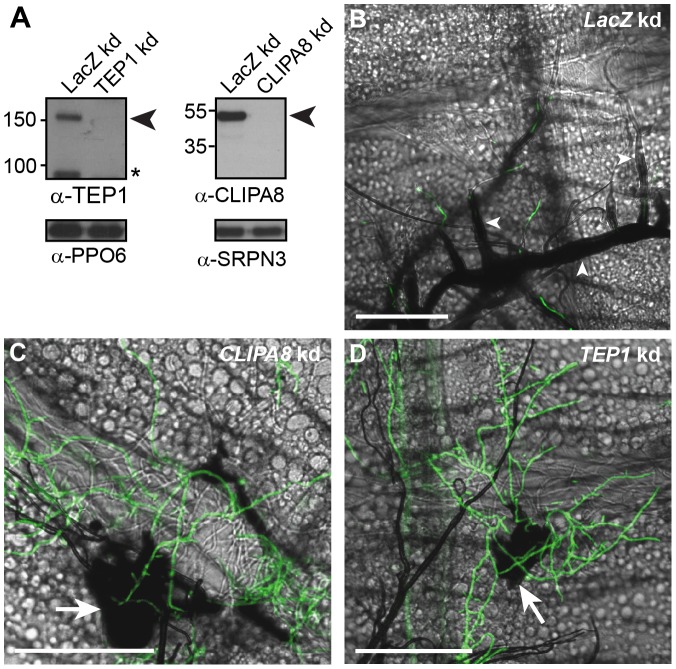
Figure 2. TEP1 and CLIPA8 are required for the melanization of hyphae.
(A) Western blot analysis showing CLIPA8 and TEP1 depletion in the hemolymph four days after silencing their corresponding genes by RNAi. Left, arrowhead indicates full-length TEP1 (TEP1-F) and asterisk denote the cleaved form of TEP1 (TEP1-C); Right, arrowhead indicates full length CLIPA8. PPO6 and SRPN3 were used as loading controls. (B) Abdomens dissected from LacZ kd mosquitoes at 48 h post-injection of 200 conidia of GFP-expressing B. bassiana (Green), show intense melanization of hyphae (arrowheads) (C and D) Absence of melanin around hyphae in CLIPA8 and TEP1 kd mosquitoes, respectively. Note, however, the strong melanization associated with the base of the mycelium in each of these genotypes (arrows). Scale bars are 100 µm.

