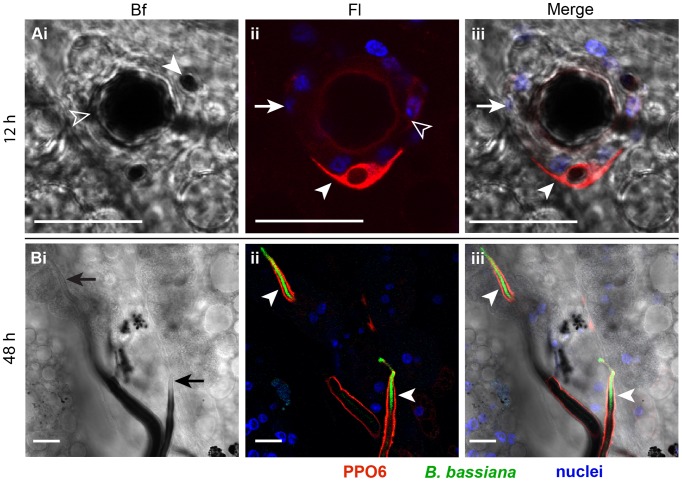
Figure 3. B. bassiana infection triggered cellular and humoral melanotic responses in the mosquito.
Abdomens were dissected at (A) 12 and (B) 48 h post-injection of each mosquito with 200 conidia of GFP-expressing B. bassiana and immunostained with α-PPO6 antibody. Shown are bright field (Bf), fluorescence (Fl), and merged bright field and fluorescence (Merge) images of confocal sections. (A) PPO staining of a germinating conidium. A conidium germinating despite being melanized (Ai, open arrowhead) exhibited an enlarged size compared to a nearby non-germinating spore (Ai, filled arrowhead). A cellular layer of hemocytes surrounding the germinating conidium (Aii and Aiii, arrows). These hemocytes exhibited no or faint PPO signal (Aii, open arrowhead). A hemocyte expressing PPO (Aii and Aiii, filled arrowhead) was resting on top of the cells that surrounded the germinating conidium. (B) PPO staining of hyphae. The apical parts of hyphal filaments exhibited a thin melanin coat (Bi, arrows) but strong PPO signal (Bii and Biii, arrowheads). The absence of hemocytes around these hyphae inform a humoral melanotic response. PPO6 (red), GFP-expressing B. bassiana (green), nuclei (blue). All scale bars are 20 µm.

