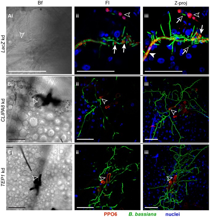
Figure 5. PPO recruitment to hyphae requires TEP1 and CLIPA8.
Mosquito abdomens were dissected from (A) LacZ, (B) CLIPA8 and (C) TEP1 kd mosquitoes at 48 h post-injection of each with 200 conidia of GFP-expressing B. bassiana and stained with PPO6 antibody. Shown are Bright field (Bf) and fluorescence (Fl) images of confocal sections, and Z projections (Z-proj) of whole stacks. (A) An abdomen from LacZ kd mosquitoes at 100× magnification showed uniform PPO staining along an established hypha (Aiii, filled arrowhead) elaborating new hyphae and hyphal bodies. Note that melanin was barely detectable on this hyphal surface (Ai, open arrowhead) despite its intense PPO staining. A strong PPO signal was also observed at the branching points of the established hypha (Aii and Aiii, arrows with filled heads). The hyphal bodies detected were not labelled with PPO (Aii and Aiii, arrows with open heads). Shown also are PPO-expressing hemocytes (Aii and Aiii, open arrowheads). (B) CLIPA8 and (C) TEP1 kd mosquito abdomens at 40× magnification showing the absence of PPO and hence melanin from hyphal surfaces despite the extensive mycelial growth in these genotypes; melanin formation (Bi and Ci, open arrowheads) and PPO staining (Bii–iii and Cii–iii, open arrowheads) were restricted only to the base of the mycelium from which hyphae emerged. PPO6 (red), B. bassiana-GFP (green) and nuclei (blue). All scale bars are 50 µm.

