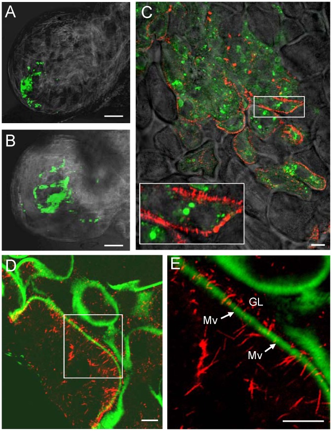
Figure 3. Pns10 tubules on actin-based microvilli of filter chamber in viruliferous leafhoppers.
At 2-day (A) or 3-day (B–E) padp, leafhopper organs were immunolabeled for Pns10 tubules with Pns10-rhodamine (red), for RDV virions with virus-FITC (green), and for actin-based microvilli with FITC-phalloidin (green), then examined by confocal microscopy. (A, B) Fluorescence micrograph of filter chamber showing green fluorescence (virus antigens) with background visualized by transmitted light. (C) Image of filter chamber merged with images with green fluorescence (virus antigens), red fluorescence (Pns10 tubules) and background visualized by transmitted light. Inset indicates an enlarged image of the boxed area. (D) Image of filter chamber merged with image of green fluorescence (actin) and of red fluorescence (Pns10 tubules). (E) Enlarged image of boxed area in panel D. GL, gut lumen. Mv, microvilli. Images are representative of multiple experiments with multiple preparations. Bars, 350 µm (A–B) and 10 µm (C–E).

