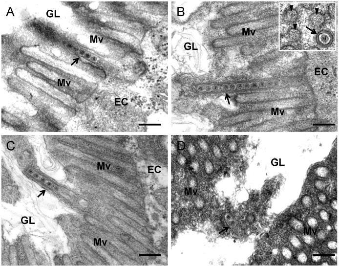
Figure 5. Transmission electron micrographs showing the association of virus-containing tubules with microvilli of anterior midgut in viruliferous leafhoppers.
(A) Closed-end of tubule (arrow) inserted into a microvillus. (B) Closed-end tubule (arrow) in contact with the inner side of distal end of microvillus. Inset, transverse section of microvillus of about 100 nm in diameter, with a virus-containing tubule (arrow) inside. Arrowheads indicate actin filaments within the microvillus. (C) Elongated tubule-associated microvillus (arrow) has formed membrane protrusion toward the lumen. (D) Tubule (arrow) in the lumen. EC, epithelial cell. GL, gut lumen. Mv, microvilli. Bars, 200 nm.

