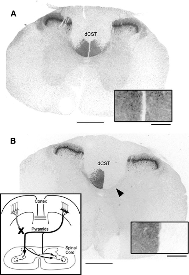
Figure 1.
Pyramidal tract lesion. Thoracic spinal cord sections showing PKC-γ immunoreactivity in sham-operated rat (A) and after unilateral pyramidal tract lesion (B). The arrowhead points to loss of staining in the contralateral dCST. The insets in A and B show dCST staining at higher magnification. The schematic in B shows lesion and anatomical organization of spared CST. Scale bars: A, B, 1000 μm; insets, 200 μm.

