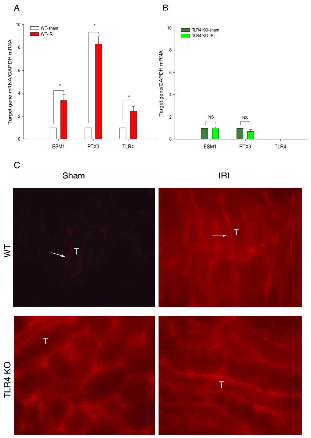
Figure 3.
TLR4 is required for increased PTX3 in AKI. Renal pedicles of WT B10 and TLR4 KO mice were clamped for 23min and kidneys harvested at 4hr reperfusion. The genes of interest (PTX3, Esm1, and TLR4) were determined by qRT-PCR and analyzed by the comparative Ct method. The calibrator gene is gene of interest taken from the sham kidney. (A) WT kidneys. (B) TLR4 KO kidneys. Error bars show mean±SEM, n= 6 in each group, *P < 0.01 IRI compared to sham; NS: not significant. (C) Immunohistology shows increased PTX3 in WT kidneys. at 4hr reperfusion. A rat anti-mouse PTX3 monoclonal antibody was used to stain frozen sections from PFA fixed tissues. Exactly the same staining conditions and exposures were used to compare PTX3 expression in sham and AKI kidneys. PTX3 is located on peritubular capillaries of the OM. PTX3 was increased on WT ischemic kidneys. No increased endothelial PTX3 was found on ischemic TLR4 KO kidneys.(X40). Arrows indicate some of many capillaries positive for PTX3; “T” indicates a few of many tubules.

