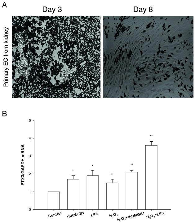
Figure 6.
Primary culture of renal EC. (A) Images of CD31+ Dynabead-isolated cells from kidney: Primary EC grown in vitro on Day 3, primary EC at confluence on Day 8. Cells showed typical cobblestone morphology. (B) PTX3 mRNA on primary EC was upregulated in vitro by H2O2(100 μM), and/or rhHMGB1(5μg/mL), and/or LPS(5μg/mL). Error bars show mean ± SEM, n=4, *P < 0.05, **P < 0.001 versus control.

