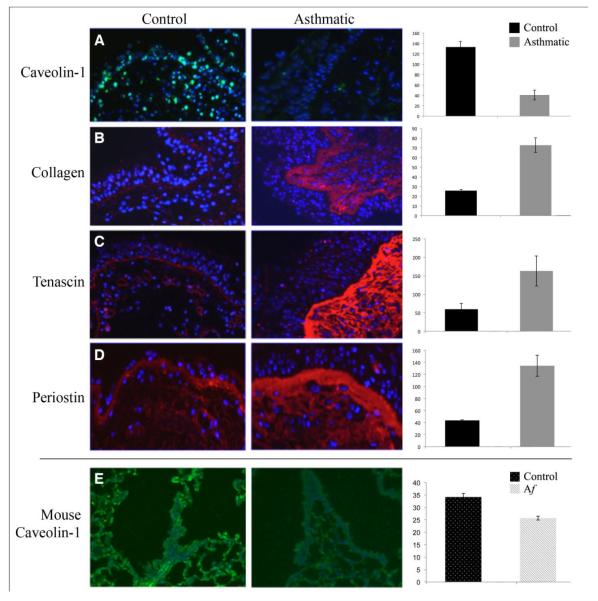
Figure 1. Caveolin-1 and ECM protein expression in human asthma lung tissue and in a murine model of allergic asthma.
A-D: Representative images of GMA-embedded human endobronchial lung biopsy sections from Control and Asthmatic subjects were stained with anti-cav-1 (A), anti-collagen (B), anti-tenascin (C) and anti-periostin (D), followed by appropriate secondary antibodies (green for A; red for B-D) and counterstained with the nuclear stain DAPI (blue). E: Representative images of anti-cav-1 stained formalin-fixed, paraffin-embedded sections from saline sensitized and challenged mice (left) and Af sensitized and challenged mice (right). In A through E, staining intensity was quantified by densitometry using Image J1.32 NIH software (five images per human subject, four subjects in each category). Average staining intensity ± s.e.m. is expressed in arbitrary units and shown to the right of each set of micrographs. The statistical significance of these data was determined using Student’s t-test. Key: Af: Aspergillus fumigatus.

