
Figure 1.
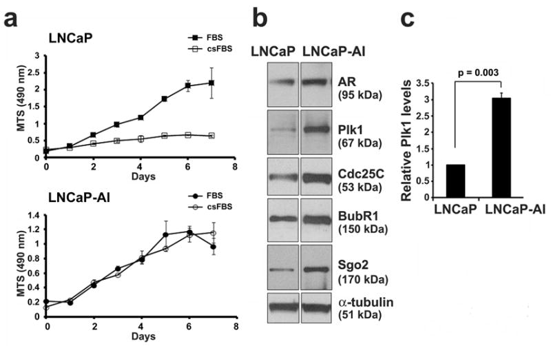
The mitotic kinase Plk1 is overexpressed in LNCaP-AI cells. (a) LNCaP and LNCaP-AI PCa cells were cultured in full media (FBS) or androgen-depleted media (csFBS). Cell growth over 7 days was determined by an MTS (OD 490 nm) growth assay. Results from triplicate samples (mean ± SD) in a representative experiment from n = 3 independent experiments are shown. (b) Lysates (20 μg) from LNCaP and LNCaP-AI cells grown in full media were immunoblotted for the androgen receptor (AR) and mitotic proteins as shown. α-tubulin was used as a loading control. (c) Relative Plk1 protein levels were determined (Plk1/tubulin and normalized against the value in LNCaP cells) (mean ± SD, n = 3 experiments).